

Breast WTA HistoSeg + LazySlide image features#
Overview#
This tutorial documents the upgraded RNA + HistoSeg structure + H&E image
workflow for the Atera breast WTA Xenium sample. HistoSeg provides tissue
structure contours, LazySlide provides WSI tile embeddings and vision-language
prompt-similarity scores when the selected model supports text embeddings, and
pyXenium.multimodal aggregates those outputs into structure-level image/RNA
association tables.
The pyXenium side is intentionally a thin integration layer. It does not run HistoSeg segmentation and it does not vendor LazySlide image-model code.
Biological question#
For each HistoSeg structure in the breast WTA sample:
Which foundation-model image features distinguish this structure from the other structures?
Which PLIP/CONCH/OmiCLIP prompt terms are enriched in the tiles assigned to the structure?
Do structure-level H&E features align with Xenium RNA programs and boundary hypotheses?
Workflow boundary#
Package |
Responsibility |
|---|---|
HistoSeg |
Structure segmentation, contour/ROI GeoJSON, mask QC |
LazySlide |
WSI opening, H&E tiling, pathology foundation model feature extraction, optional vision-language prompt scoring, spatial tile domains |
pyXenium |
Xenium/H&E alignment, tile-to-structure assignment, structure aggregation, RNA/image association |
Python API#
from pyXenium.multimodal import run_histoseg_lazyslide_structure_workflow

result = run_histoseg_lazyslide_structure_workflow(
    "/path/to/WTA_Preview_FFPE_Breast_Cancer_outs",
    contour_geojson="/path/to/xenium_explorer_annotations.s1_s5.generated.geojson",
    contour_key="histoseg_structures",
    output_dir="/path/to/a100/run",
    he_source_path="/path/to/WTA_Preview_FFPE_Breast_Cancer_he_image.tiffslide_pyramid.tif",
    wsi_reader="tiffslide",
    model="plip",
    text_model="plip",
    tile_px=224,
    mpp=0.5,
    device="cuda",
    batch_size=64,
    table_format="parquet",
)
model is the LazySlide image/foundation model used for tile embeddings. It can
be any model supported by the local LazySlide installation, for example plip,
conch, uni, uni2, gigapath, virchow, or related model-zoo entries.
text_model is separate and is only used for tile-level prompt scoring. It must
share the same image-text latent space as the image model. In practice,
text_model="plip" is valid with model="plip" and text_model="conch" is
valid with model="conch". Vision-only encoders such as UNI, Virchow, or
GigaPath produce embeddings and spatial domains but do not automatically assign
pathology names. Use text_model="none" to disable prompt scoring explicitly.
The workflow writes:
image_contours.parquet
tile_features.parquet
tile_assignments.parquet
structure_image_features.parquet
structure_differential_features.parquet
structure_rna_summary.parquet
structure_program_scores.parquet
contour_multimodal_summary.parquet
contour_image_molecular_associations.parquet
wta_pathway_partial_correlations.parquet
molecular_prediction_benchmark.parquet
morphomolecular_hero_targets.parquet
morphomolecular_hero_contours.parquet
rna_image_associations.parquet
program_image_associations.parquet
run_manifest.json
A100 run#
Use the A100 runner in benchmarking/lazyslide_a100/:
export PYXENIUM_REPO=/path/to/pyXenium
export A100_ENV_DIR=/path/to/envs/pyxenium-lazyslide
bash benchmarking/lazyslide_a100/scripts/bootstrap_a100_env.sh

export PYXENIUM_ATERA_DATASET=/path/to/WTA_Preview_FFPE_Breast_Cancer_outs
export HISTOSEG_GEOJSON=/path/to/xenium_explorer_annotations.s1_s5.generated.geojson
export A100_OUTPUT_DIR=/path/to/runs/histoseg_lazyslide_breast_wta_plip
export LAZYSLIDE_MODEL=plip
export LAZYSLIDE_TEXT_MODEL=plip

bash benchmarking/lazyslide_a100/scripts/run_a100_histoseg_lazyslide.sh \
  --max-tiles 2000
WSI preparation#
The original Atera breast WTA OME-TIFF stores RGB as planar SYX. tifffile
can see the internal levels, but WSIData/open_wsi defaults to OpenSlide and
only recognizes one level for this file layout. That made the first direct WSI
attempt behave like a one-level 17 GB image.
The fix is to rewrite the H&E image once into a tiffslide-readable tiled
pyramidal BigTIFF with interleaved YXS RGB:
PYTHONPATH=src \
/data/taobo.hu/pyxenium_lazyslide_breast_wta_20260507/envs/plip-patch/bin/python \
  benchmarking/lazyslide_a100/scripts/prepare_tiffslide_pyramid.py \
  --input /data/taobo.hu/pyxenium_lazyslide_breast_wta_20260507/data/WTA_Preview_FFPE_Breast_Cancer_he_image.ome.tif \
  --output /data/taobo.hu/pyxenium_lazyslide_breast_wta_20260507/data/WTA_Preview_FFPE_Breast_Cancer_he_image.tiffslide_pyramid.tif \
  --tile-px 512 \
  --jpeg-quality 90 \
  --verify
The prepared WSI validates as 10 levels with MPP 0.2738 and is recorded in
prepared_wsi_manifest.json.
The full direct LazySlide PLIP command used for this RTD snapshot was:
CUDA_VISIBLE_DEVICES=7 \
/data/taobo.hu/pyxenium_lazyslide_breast_wta_20260507/envs/plip-patch/bin/python \
  benchmarking/lazyslide_a100/scripts/run_histoseg_lazyslide_workflow.py \
  --dataset-root /data/taobo.hu/pyxenium_lr_benchmark_2026-04/data/source_cache/breast/WTA_Preview_FFPE_Breast_Cancer_outs/spatialdata.zarr \
  --histoseg-geojson /data/taobo.hu/pyxenium_lazyslide_breast_wta_20260507/data/xenium_explorer_annotations.s1_s5.generated.geojson \
  --contour-id-key name \
  --he-source-path /data/taobo.hu/pyxenium_lazyslide_breast_wta_20260507/data/WTA_Preview_FFPE_Breast_Cancer_he_image.tiffslide_pyramid.tif \
  --wsi-reader tiffslide \
  --output-dir /data/taobo.hu/pyxenium_lazyslide_breast_wta_20260507/runs/direct_lazyslide_plip_full_text \
  --model plip \
  --text-model plip \
  --batch-size 64 \
  --table-format parquet
A100 PLIP result snapshot#
The committed RTD artifacts in this page come from a completed direct WSI LazySlide run on GPU 7 using PLIP.
Field |
Value |
|---|---|
Workflow |
|
WSI reader |
|
LazySlide |
|
GPU |
NVIDIA A100-SXM4-40GB |
Torch |
|
Embedding model |
|
Prompt scoring model |
|
HistoSeg contours |
1,578 |
LazySlide tiles |
3,115 |
Assigned tiles |
3,114 |
HistoSeg structures |
5 |
Embedding dimensions |
512 |
Runtime |
3,989.3 seconds |
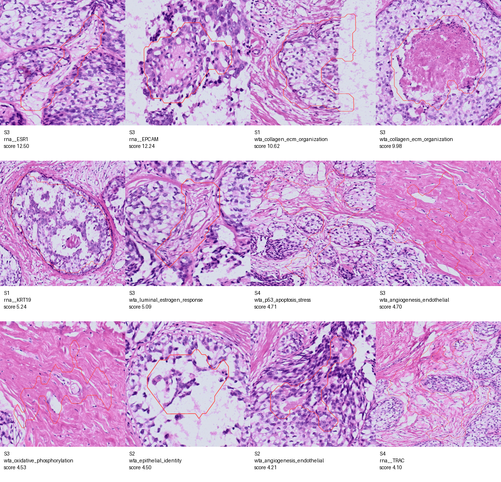
The PLIP prompt terms used for zero-shot image-text scoring were:
ductal epithelium
invasive carcinoma
in situ carcinoma
fibrotic stroma
immune infiltrate
necrosis
adipose tissue
vascular stroma
lumen or secretion
These terms are a manually curated breast histology prompt set
(breast_histology_v1). They are not HistoSeg structure names and they are not
pathologist-confirmed diagnostic labels.
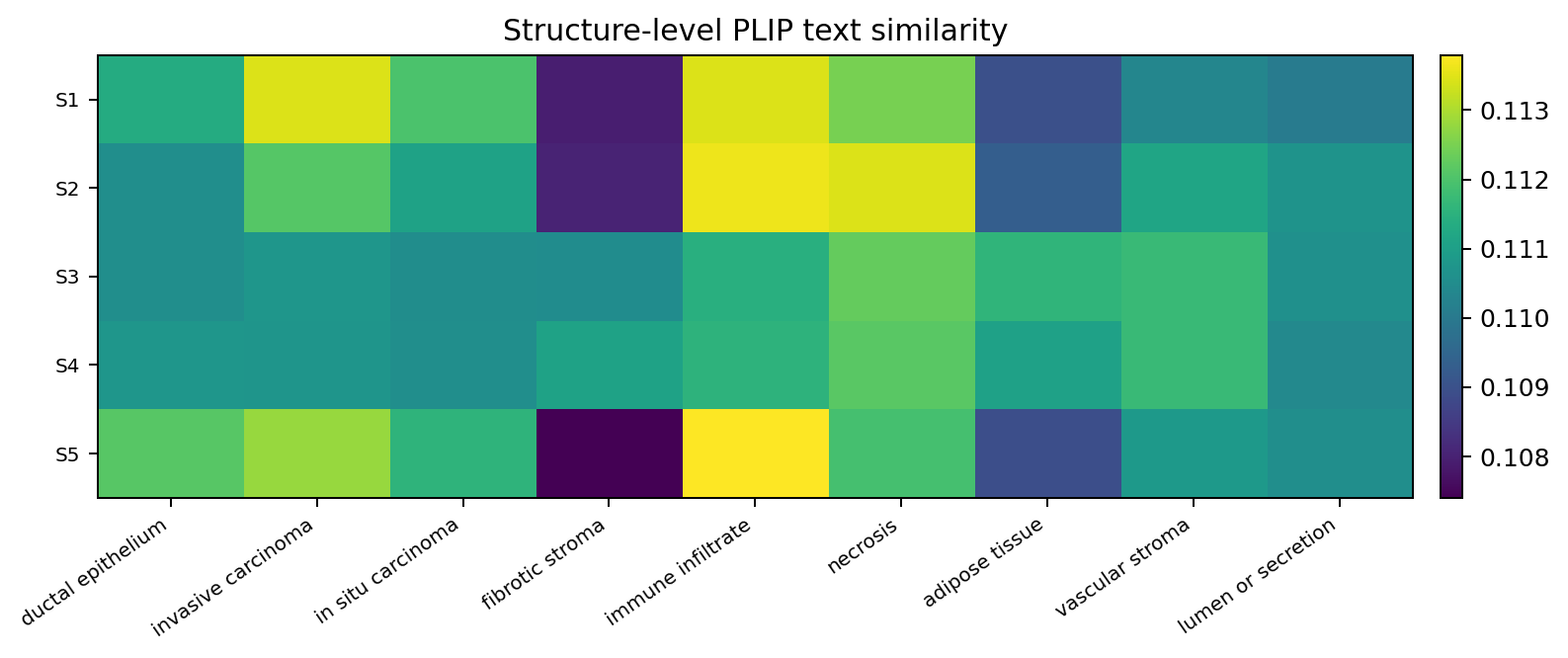
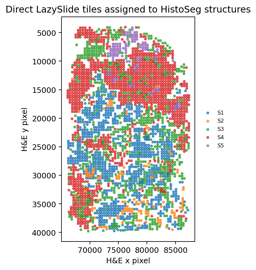
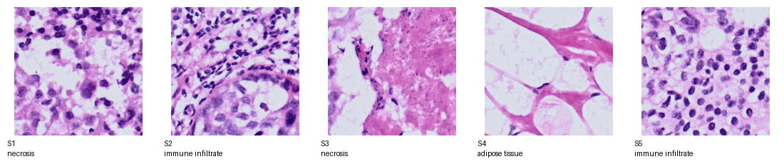
Structure-level prompt scores#
HistoSeg structure |
Tiles |
Top mean PLIP prompt |
Top tile prompt mode |
Enriched prompt-similarity terms |
|---|---|---|---|---|
S1 |
866 |
invasive carcinoma |
immune infiltrate |
invasive carcinoma, immune infiltrate, in situ carcinoma |
S2 |
140 |
immune infiltrate |
immune infiltrate |
immune infiltrate, necrosis, invasive carcinoma |
S3 |
797 |
necrosis |
necrosis |
adipose tissue, vascular stroma, fibrotic stroma |
S4 |
1,184 |
necrosis |
fibrotic stroma |
fibrotic stroma, vascular stroma, adipose tissue |
S5 |
127 |
immune infiltrate |
immune infiltrate |
invasive carcinoma, immune infiltrate, in situ carcinoma |
The enriched terms are one-vs-rest positive PLIP prompt-similarity features with FDR < 0.05 when available. They should be interpreted as image-language features, not as diagnostic labels. The score range is narrow, so the most useful signal is the structure-to-structure contrast rather than the absolute name of a single top prompt.
Contour-scale WTA morphomolecular programs#
The WTA downstream analysis reuses the completed direct PLIP tile run and scores breast/TME gene programs from the Xenium whole-transcriptome matrix at HistoSeg-contour scale. The key test is no longer whether H&E beats structure labels in cross-validated prediction. In this single sample, HistoSeg labels explain much of the coarse molecular variance. The stronger result is residual: after controlling for HistoSeg structure, contour centroid, and cell-to-boundary distance, prompt-independent PLIP embedding axes remain associated with WTA program activity.
Top residual WTA program associations:
Rank |
WTA program |
Best H&E axis |
Partial Spearman rho |
FDR |
|---|---|---|---|---|
1 |
luminal_estrogen_response |
|
-0.597 |
8.64e-25 |
2 |
epithelial_identity |
|
-0.496 |
1.10e-16 |
3 |
basal_squamous_state |
|
0.492 |
1.89e-16 |
4 |
unfolded_protein_response |
|
0.491 |
2.30e-16 |
5 |
oxidative_phosphorylation |
|
0.485 |
6.58e-16 |
6 |
p53_apoptosis_stress |
|
-0.439 |
8.12e-13 |
7 |
tls_b_cell_plasma |
|
-0.409 |
4.57e-11 |
8 |
cell_cycle_proliferation |
|
0.398 |
1.97e-10 |
9 |
her2_amplicon_signaling |
|
0.389 |
5.73e-10 |
10 |
t_cell_exhaustion_checkpoint |
|
-0.377 |
2.52e-09 |
The complete table is available as
wta_pathway_partial_correlations.csv.
The hero contour tables are available as
morphomolecular_hero_targets.csv
and
morphomolecular_hero_contours.csv.
Visual outputs#
Artifact files#
The copied A100 snapshot is stored in:
docs/_static/tutorials/multimodal_histoseg_lazyslide_breast_wta/
Key files are:
Interpretation rules#
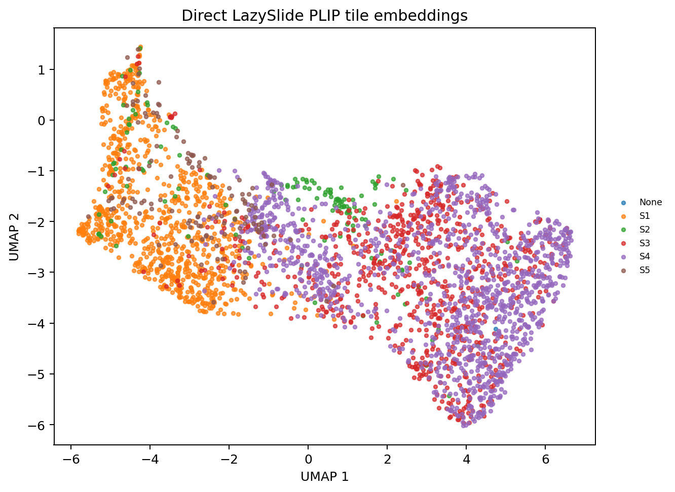
structure_image_featuresis the main table for asking what image signatures each HistoSeg structure carries.structure_differential_featuresranks one-vs-rest image features per structure.The current RTD snapshot reports direct LazySlide WSI tiling, PLIP image embeddings, PLIP prompt-similarity scores, structure-level RNA summaries, contour-level WTA program scores, partial morphomolecular associations, and program/image associations.
In the Breast WTA single-sample snapshot,
structure_onlyremains the strongest cross-validated baseline for many epithelial markers and programs. Treat the WTA result as evidence for within-structure residual molecular decoding, not as a claim that H&E embeddings improve every molecular prediction benchmark beyond HistoSeg labels.PLIP is the first required A100 result. CONCH, OmiCLIP, UNI, Virchow, GigaPath, and other LazySlide foundation models can be run through the same
modelentry point when the model files, licenses, and credentials are available. Vision-only models contribute embeddings; vision-language models can also contribute prompt scores.
Current implementation status#
The pyXenium API, optional dependency boundary, A100 runner, WSI preparation script, direct LazySlide WSI backend, artifact schema, and full A100 PLIP image-feature snapshot are implemented. The earlier patch-corpus fallback remains useful for quick checks, but this page now reports the direct WSI LazySlide result.